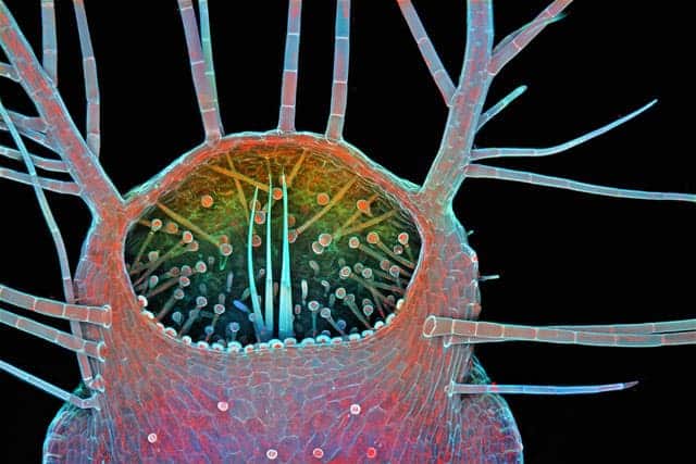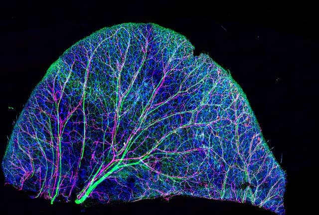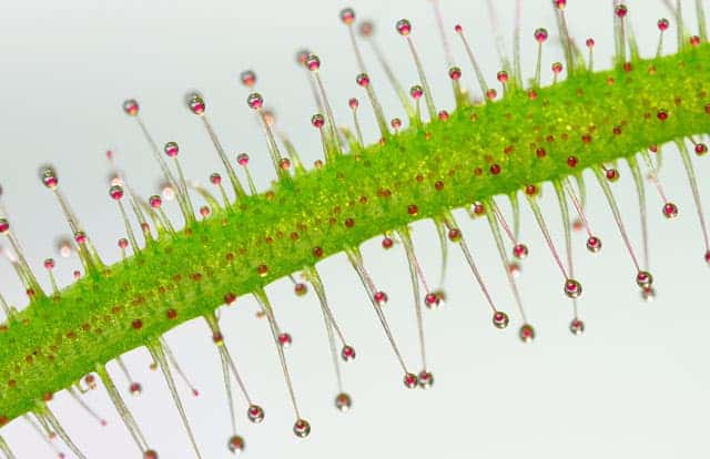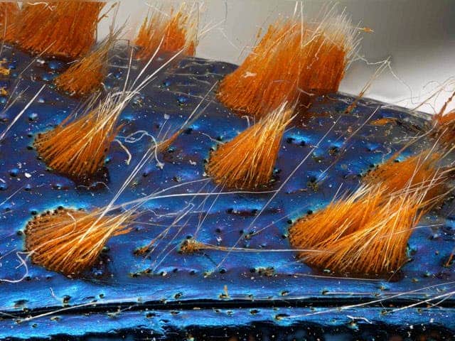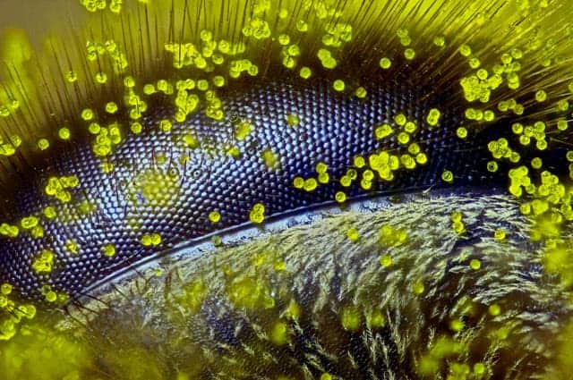
Every year Nikon holds the Small World Photomicrography Competition awarding the very finest photographers that capture the essence of the micro world. For 2015, the winner was Ralph Grimm from Australia with a close-up of a bee eye covered in dandelion pollen grains. This was no lucky shot. Grimm says it took four hours to prepare, including setting focus increments, proper illumination and stacking.
“In a way I feel as though this gives us a glimpse of the world through the eye of a bee,” says Grimm. “It’s a subject of great sculptural beauty, but also a warning- that we should stay connected to our planet, listen to the little creatures like bees, and find a way to protect the earth that we all call home.”
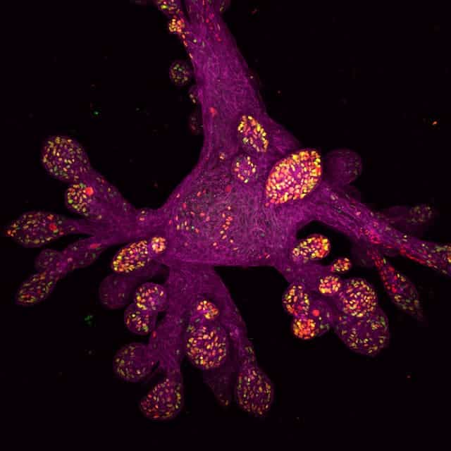
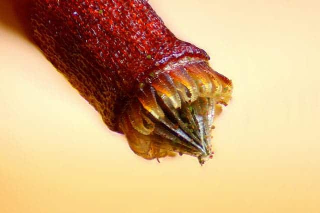
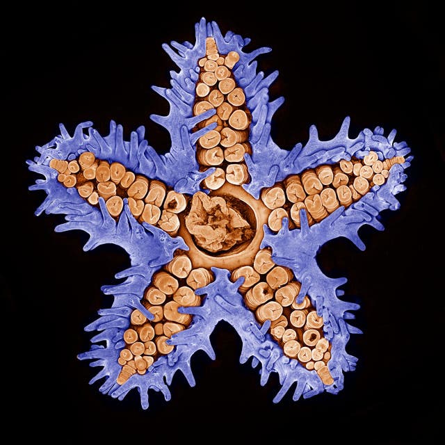
“Each year we are blown away by the incredible quality and quantity of microscopic images submitted from all over the world, from scientists, artists, and photomicrographers of all levels and backgrounds. This year was certainly no exception,” said Eric Flem, Communications Manager, Nikon Instruments. “Judges had their work cut out for them in narrowing down from such a rich pool of applicants, and we are so pleased with the results. Each of these winning images exhibits the exemplary technique, scientific discipline and artistry for which Nikon Small World is known.